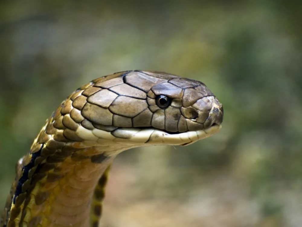
Reptile scales are marvels of evolutionary engineering. Far from being static, inert armor plates, these complex epidermal structures vary vastly across taxa, tailored precisely to the ecological niches of their owners. Varanids and crocodilians possess hard osteoderms—bone embedded directly within their scales—while rattlesnakes feature keeled scales designed for sensory and locomotion benefits. The skin of Uromastyx and chuckwallas is remarkably elastic to accommodate inflation behaviors used to deter predators or regulate social status. In many geckos and all snakes, the eyelids have fused and cleared to form the spectacle, a transparent scale protecting the eye.
For every environmental challenge a reptile faces, its scaly skin offers profound evolutionary advantages. Yet, because scales and skin are so fundamentally tied to a reptile’s overall physiological well-being, dermatologic disruptions can quickly cascade into life-threatening systemic crises.
To help reptile keepers, conservationists, and veterinary professionals better understand the complex landscape of reptilian dermatology, this report examines the broad spectrum of infectious and non-infectious skin pathologies, their clinical implications, and the essential protocols required for prevention and veterinary intervention.
(Disclaimer: This article is intended for educational purposes and does not serve as a diagnostic or therapeutic manual. Any observed skin abnormalities should be evaluated promptly by a qualified reptile or exotic animal veterinarian.)
Part I: The Main Facts — Understanding Reptile Epidermal Anatomy and Pathophysiology
The reptilian integumentary system serves as the primary barrier against physical trauma, desiccation, and opportunistic pathogens. Unlike mammalian skin, which heals through extensive soft-tissue remodeling and liquid purulence, reptilian skin responds to injury and infection through distinct physiological mechanisms.
Infectious Dermatologic Landscapes
When pathogens breach the epidermal barrier, reptiles frequently develop infectious dermatitis or localized abscessation. Because reptiles lack peroxidase enzymes—the biochemical components responsible for liquefying pus in mammals—their abscesses do not produce watery discharge. Instead, reptile pus takes on a characteristic caseous (cheesy), semi-solid consistency. This biological quirk dictates that standard mammalian treatment protocols often fail; simple lancing and draining are insufficient. Thick-walled abscesses typically require surgical excision because the fibrous walls that contain the infection also prevent systemic antibiotics from penetrating the core of the lesion.
Non-Infectious Stressors and Husbandry Failures
A significant percentage of dermatologic presentations in captivity stem from suboptimal husbandry. Environmental factors—most notably inappropriate thermal gradients, improper relative humidity, inadequate ultraviolet (UV) lighting, and poor substrate hygiene—account for the majority of non-infectious skin diseases. When the delicate balance of a captive ecosystem fails, the skin is often the first organ system to reflect the systemic stress.
Part II: Chronology of Disease Progression — How Minor Husbandry Flaws Escalate into Systemic Crises
To understand how skin diseases develop in captive reptiles, it is helpful to trace the chronological evolution of a typical dermatologic failure, such as improper shedding (dysecdysis) or localized trauma, and observe how it progresses if left unmanaged.
Stage 1: The Initial Insult or Environmental Fluctuation
The process often begins subtly. For instance, an enclosure’s relative humidity drops below acceptable thresholds due to seasonal weather changes or improper misting schedules. Alternatively, a reptile sustains a minor abrasion from a poorly secured enclosure decoration or a cage mate. At this microscopic level, the epidermal barrier is compromised.
Stage 2: Epidermal Dysfunction and Pathogen Infiltration
In the case of humidity drops, the natural ecdysis (shedding) cycle is interrupted. Old epidermal layers fail to separate cleanly from the new skin beneath. This results in "stuck shed," particularly around vulnerable extremities like digits and the tail tip. Concurrently, micro-abrasions or areas of retained shed become prime real estate for opportunistic pathogens, such as bacteria (Pseudomonas spp. or Devrisea agamarum) or fungal spores (such as snake fungal disease agents or Chrysosporium anamorph of Nannizziopsis vriesii, commonly known as yellow fungus).
Stage 3: Localized Necrosis and Inflammation
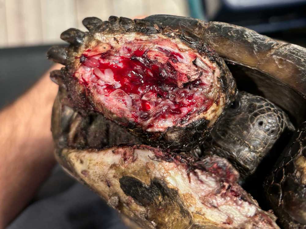
As pathogens colonize the compromised tissue, local inflammation ensues. In cases of chronic stuck shed, layers of retained skin constrict blood flow to the underlying digits or tail tips. Over days to weeks, this ischemic environment leads to tissue death (necrosis). If a bacterial infection takes hold, clinical signs such as ulceration, hemorrhaging, crusting, and localized pain upon palpation become apparent. This stage frequently manifests colloquially as "rot" (e.g., tail rot or scale rot).
Stage 4: Systemic Dissemination and Organ Failure
If the underlying husbandry is not corrected and veterinary intervention is delayed, localized dermatologic diseases routinely progress to systemic infections. Bacterial toxins, fungal hyphae, or inflammatory byproducts enter the bloodstream. Septicemia, metabolic bone disease, renal failure, or hepatic lipidosis can rapidly develop. At this advanced stage, the prognosis drops significantly, requiring aggressive, long-term medical and surgical intervention to save the animal’s life.
Part III: Supporting Data & Classification of Reptile Skin Pathologies
Dermatologic diseases in reptiles are broadly categorized into infectious and non-infectious etiologies. Below is an exhaustive breakdown of the primary conditions encountered in veterinary practice.
1. Infectious Dermatologic Diseases
Bacterial Pathogens
- Bacterial Dermatitis: Frequently presents as ulceration, bleeding, scale loss, and crusting. A notable pathogen is Devrisea agamarum, which heavily impacts Uromastyx and agamid lizards. Notably, bearded dragons (Pogona vitticeps) often act as asymptomatic carriers—appearing entirely healthy while shedding the pathogen to highly susceptible cage mates.
- Scale Rot and "X Rot": Generalized or localized bacterial infections affecting the ventral scales, tail, or body. These conditions are typically secondary to filthy, damp substrate and poor water quality, culminating in necrotic tissue loss.
- SCUD (Septic Cutaneous Ulcerative Dermatitis): Common in aquatic and semi-aquatic chelonians (turtles and tortoises), this condition begins as localized shell and skin ulcers but rapidly progresses into a systemic, life-threatening infection.
Viral Pathogens
- Poxviruses: Affect a diverse array of reptiles, exhibiting pathogenic behaviors comparable to mammalian poxvirus infections, leading to proliferative skin lesions.
- Fibropapillomatosis: Highly prevalent in sea turtles, caused by chelonid herpesviruses that induce massive, debilitating tumor-like nodules on the soft tissues, eyes, and flippers.
- Ranaviruses and Papillomaviruses: Increasingly recognized agents that cause systemic illnesses accompanied by significant skin lesions and epidermal thickening.
Fungal Pathogens
- Yellow Fungus Disease: Caused by Chrysosporium species, this highly contagious and aggressive fungal infection affects bearded dragons and other lizards, frequently proving fatal if it invades deep tissues and internal organs.
- Snake Fungal Disease (SFD): Caused by Ophidiomyces ophiodiicola, this pathogen targets wild and captive snakes, causing crusty scabs, discolored scales, and facial disfigurement.
- Emydomyces fungal infections: Target box turtles and pond turtles, leading to severe shell and skin degradation.
Parasitic Infestations
- Ectoparasites: Snake mites (Ophionyssus natricis) are rampant in captive collections. They feed on blood, cause severe anemia, create micro-abrasions that invite secondary bacterial/fungal infections, and act as vectors for viral diseases.
- Endoparasites: Certain internal parasites (such as tapeworms and nematodes) utilize reptiles as intermediate hosts, encapsulating within the tissues to form subcutaneous nodules or granulomas. This biological strategy ensures that when a predator consumes the reptile, the parasite successfully reaches the primary host’s gastrointestinal tract.
2. Non-Infectious Dermatologic Diseases
Dysecdysis (Abnormal Shedding)
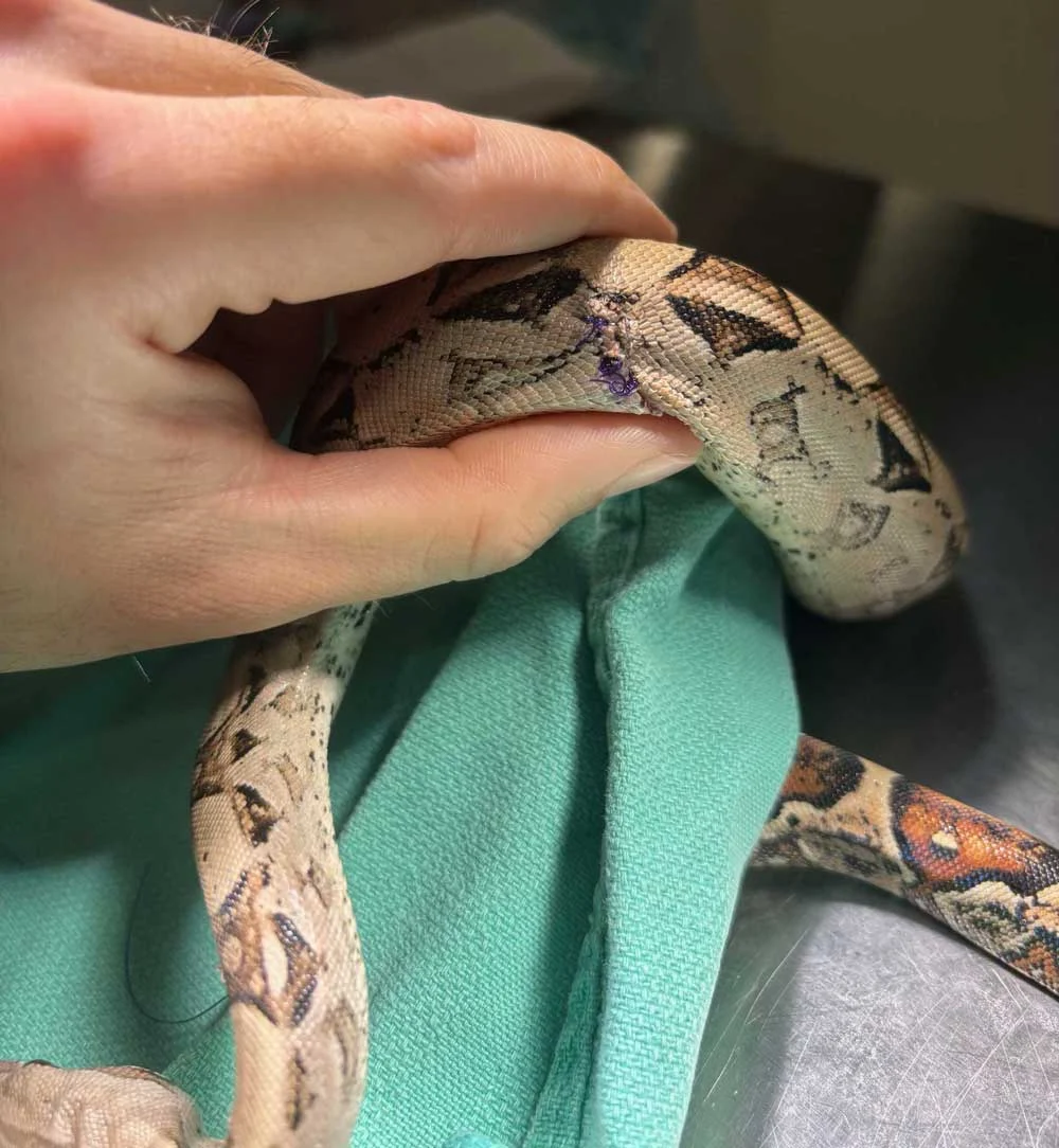
Driven by low humidity, poor nutrition, or lack of proper rough surfaces to rub against during shedding. While mild cases respond to warm water soaks and gentle manual removal, severe chronic cases result in multi-layer constriction, digital necrosis, and necessary surgical amputations. Conversely, accelerated shedding can be triggered by skin irritation, mites, surgical sutures, or underlying metabolic disturbances.
Trauma and Mechanical Injury
Caused by unstable cage decor, improper handling (e.g., giant day geckos sloughing skin in defense), or encounters with cage mates and prey items. Rare presentations include body wall hernias—observed in leopard geckos—where internal organs rupture through a tear in the abdominal wall and push outward beneath the intact skin.
Thermal Burns
Occur when reptiles lack safe basking environments. Common culprits include commercial "heat rocks" (widely discouraged by reptile veterinarians due to unreliable heating thermostats), unregulated heat mats, or undersized basking lamps that force a large animal (such as a monitor lizard) to absorb intense heat unevenly across its body.
Nutritional and Metabolic Disorders
- Vitamin A Deficiency: Critical for maintaining healthy, moist mucous membranes. A lack of Vitamin A leads to keratinous debris accumulation, causing blocked tear ducts and eye caps in leopard geckos, aural (ear) abscesses in aquatic turtles, hemipenal plugs in lizards, and stomatitis (mouth rot).
- Gout (Articular and Visceral): A metabolic disorder resulting from uric acid buildup. Articular gout causes excruciatingly painful joint swellings that yield chalky white urate deposits upon veterinary aspiration.
Neoplasia (Cancer)
Reptiles are susceptible to various malignancies. Documented cases include melanomas in turtles, squamous cell carcinomas, mast cell tumors, and sarcomas in bearded dragons, and lipomas in snakes. Not every subcutaneous mass is an abscess; definitive diagnosis requires cytology or histopathology.
Part IV: Official Veterinary Perspectives and Official Responses
To combat the rising incidence of preventable reptile skin diseases, veterinary organizations such as the Association of Reptile and Amphibian Veterinarians (ARAV) have continually updated their husbandry and clinical care guidelines.
According to avian and exotic animal clinicians, client education remains the single most effective tool in preventative medicine. Dr. Eric Los Kamp, an exotic animal and wildlife veterinarian practicing in Florida, emphasizes that skin pathologies are rarely isolated events.
"For any challenge reptiles face, their scaly skin finds ways to provide their owners advantages in adaptation. With scales so integral to their lives, when there are issues with the skin, it can cause significant issues," Dr. Los Kamp notes. "The best way to minimize skin diseases in reptiles is providing optimal thermal and humidity gradients. Substrate used should be cycled and replaced regularly, as substrate that is too wet leads directly to infections, while substrate that is too dry causes catastrophic shedding failures."
Veterinary boards also stress the danger of self-diagnosis and over-the-counter treatments. Topical human antibiotic ointments containing certain petroleum bases or steroids can severely disrupt a reptile’s delicate epidermal respiration and immune response, often worsening bacterial and fungal infections. Professional veterinary evaluation—utilizing skin scrapings, fungal cultures, biopsies, and blood panels—is mandatory for targeted, safe treatment.
Part V: Implications for Keepers, Conservationists, and the Pet Trade
The prevalence of dermatologic diseases in reptiles carries profound implications across multiple sectors of the reptile community:
- Animal Welfare and Ethics: The high rate of husbandry-related skin diseases underscores the ongoing ethical challenges of keeping exotic animals in captivity. Ensuring that a reptile’s complex microclimate needs are met requires significant financial investment, architectural knowledge, and daily diligence.
- Biosecurity and Disease Spread: As demonstrated by asymptomatic carriers of Devrisea agamarum and highly contagious pathogens like yellow fungus and snake fungal disease, lax quarantine protocols can devastate multi-animal collections, breeding facilities, and wild conservation projects.
- The Role of the Exotic Veterinarian: The veterinary medical community must continue bridging the gap between wild reptile biology and captive husbandry. By shifting the focus from reactive surgery (such as tail amputations and abscess debridement) to proactive husbandry education, the reptile community can drastically reduce the incidence of debilitating skin disorders.
Summary Prevention Checklist for Reptile Keepers
- Maintain Species-Specific Gradients: Provide accurate, well-monitored temperature zones and humidity levels utilizing high-quality digital hygrometers, thermometers, and reliable thermostats.
- Optimize Substrate Management: Spot-clean daily and perform complete substrate replacements regularly. Avoid overly damp substrates that foster bacterial proliferation, and ensure adequate moisture during shedding cycles.
- Eliminate Unsafe Heating Sources: Discard commercial heat rocks and regulate all under-tank heaters and radiant heat panels with precise thermostats.
- Enforce Strict Quarantine Protocols: Isolate new additions to a collection for a minimum of 90 days to screen for ectoparasites, fungal spores, and infectious dermatitis.
- Seek Early Veterinary Care: At the first sign of scale discoloration, retained shed, swelling, or crusting, consult an experienced exotic animal veterinarian immediately to halt disease progression before it turns systemic.