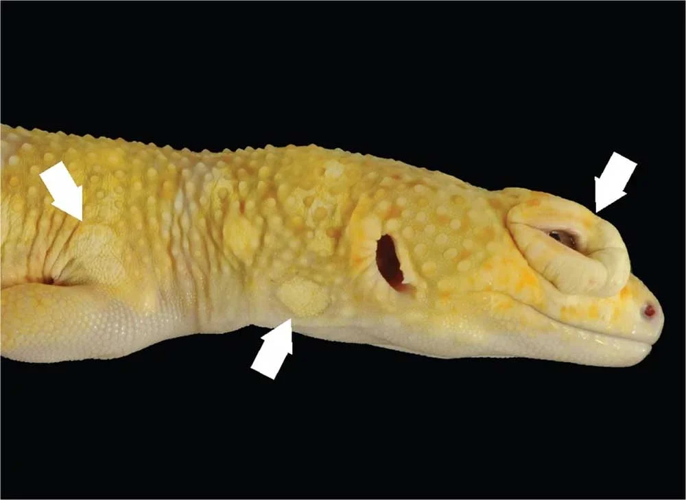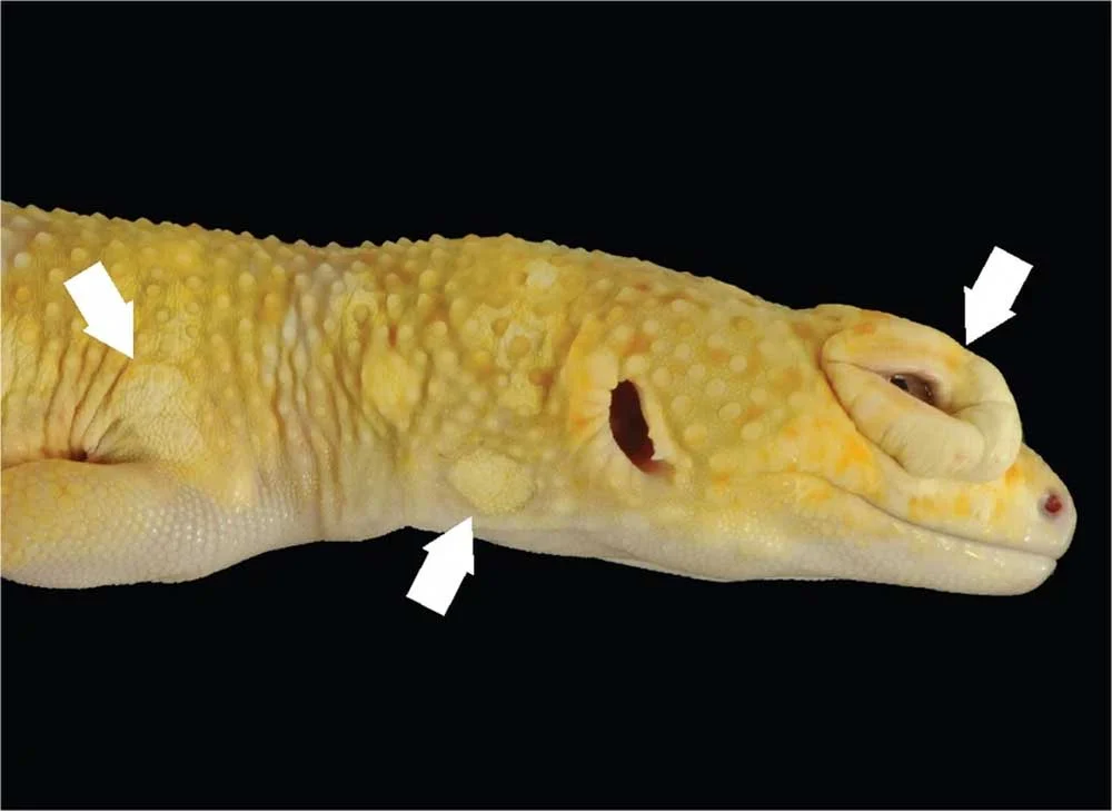
Reptile scales are marvels of evolutionary engineering. Far from being static armor, these specialized structures are dynamic, highly variable adaptations that evolve to meet the distinct ecological niches of different taxa. Varanid lizards and crocodilians feature rigid osteoderms—actual bone—embedded within their scales for enhanced protection. Rattlesnakes boast keeled scales with ridged centers that aid in traction and acoustic signaling, while Uromastyx and chuckwallas feature elastic hides that accommodate dramatic body inflation behaviors. In certain geckos and all snakes, the scales over the eyes fuse and clear to form the spectacle, a permanent protective lens replacing movable eyelids.
Because integumentary health is deeply tied to a reptile’s physiological well-being, any disruption to the skin can trigger a cascade of systemic issues. To explore the complexities of these conditions, this report examines the broad spectrum of infectious and non-infectious dermatologic diseases affecting captive reptiles, drawing on clinical insights from veterinary professionals.
(Note: This overview is strictly informational and does not serve as a diagnostic or treatment guide. Any suspected skin abnormalities in captive reptiles should be evaluated immediately by a qualified exotic animal veterinarian.)
Main Facts: The Anatomy of Reptile Skin Vulnerability
The reptilian integumentary system serves as the primary barrier against environmental pathogens, mechanical trauma, and moisture loss. However, captive environments present artificial challenges that frequently compromise this delicate barrier. Unlike mammals, reptiles cannot mount identical inflammatory or immune responses. For instance, reptiles lack peroxidase enzymes—the compounds responsible for liquefying pus in mammalian wounds. Consequently, when a reptile develops an infection, the resulting cellular accumulation is remarkably dense and caseous (cheesy or solid), changing how veterinarians must approach surgical and medical interventions.
Dermatologic diseases in reptiles are generally split into two categories: infectious and non-infectious. Infectious diseases involve pathogens such as bacteria, viruses, fungi, and external or internal parasites. Non-infectious disorders stem from husbandry failures, nutritional imbalances, environmental trauma, and genetic predispositions like cancer. Understanding the intersection between proper husbandry—such as heat, humidity, and nutrition—and dermatologic health is critical for preventing chronic or fatal complications.
Chronology: The Pathogenesis and Progression of Skin Disease
To understand how reptile skin diseases develop, veterinary medicine looks at the typical timeline of integumentary breakdown, ranging from initial environmental stressors to advanced systemic illness.
Phase 1: Environmental Stress and Barrier Compromise
The timeline often begins with suboptimal husbandry. If an enclosure lacks proper humidity gradients, a reptile enters the shedding cycle (ecdysis) unable to properly release its old stratum corneum. Simultaneously, poor hygiene—such as damp, soiled substrate or unmonitored aquatic filtration—weakens the epidermal barrier. Microscopic abrasions or constant moisture exposure macerates the skin, opening the door for opportunistic pathogens.
Phase 2: Pathogen Infiltration and Localized Lesions
Once the barrier is breached, secondary invaders take hold.
- Bacterial colonization leads to ulcerative dermatitis, marked by petechiation (small red dots), bleeding, and crusting.
- Fungal spores, such as those responsible for snake fungal disease or yellow fungus, begin to digest keratin, creating visible crusts, discoloration, and deep-seated granulomas.
- Ectoparasites, particularly snake mites, pierce the dermis to feed on blood, causing intense pruritus (itching), anemia, and focal tissue damage that invites further bacterial infection.
Phase 3: Chronic Encapsulation and Systemic Spread
Left unchecked, localized infections undergo pathological shifts. Because reptiles form solid, caseous exudates instead of liquid pus, chronic wounds quickly transform into walled-off abscesses. While this fibrous wall temporarily halts the immediate spread of bacteria, it simultaneously prevents systemic antibiotics from penetrating the core of the infection, necessitating surgical excision.
In the worst-case scenario, pathogens breach the local vascular bed, resulting in septicemia. Conditions such as Septic Cutaneous Ulcerative Dermatitis (SCUD) or systemic viral infections (like ranavirus or fibropapillomas) transition from superficial skin blemishes to multi-organ failure and death.
Supporting Data: Categories of Dermatologic Pathology
Clinical data from exotic veterinary hospitals highlights several key categories of skin pathology, distinguishing between infectious agents and environmental or systemic triggers.
Infectious Dermatologic Diseases
1. Bacterial Infections
Bacterial dermatitis presents in multiple guises, ranging from superficial scale loss to deep tissue ulceration. A notable pathogen is Devrisea agamarum, which heavily impacts Uromastyx and other agamid lizards. Alarmingly, popular pets like bearded dragons often act as asymptomatic carriers—exhibiting completely normal physical appearances while shedding the pathogen to highly susceptible tankmates.
Generalized regional infections are frequently grouped under colloquial terms like "tail rot" or "scale rot." Although the underlying bacteria vary, these syndromes consistently exhibit tissue necrosis. Bacterial dermatitis can also serve as the cutaneous manifestation of a deeper systemic disease, as seen in SCUD.
2. Viral Infections
Viral pathogens frequently manifest cutaneous lesions as part of a systemic disease process. Poxviruses affect a wide range of reptilian species, mirroring the pathological behavior of mammalian poxviruses. In marine conservation, the fibropapilloma virus—driven by a herpesvirus—causes massive, debilitating fibrovascular nodules on the faces and flippers of sea turtles. Ranaviruses and papillomaviruses are also well-documented culprits in reptilian dermal lesions.
3. Fungal Pathogens
Fungal dermatitis is notoriously contagious within captive collections and is frequently tied to systemic illness. High-profile examples include:
- Yellow Fungus Disease: Affects bearded dragons and other lizards, presenting as aggressive yellow-to-brown crusting lesions that invade deep tissues.
- Snake Fungal Disease (Ophidiomyces ophiodiicola): Causes facial crusting, disfiguration, and mortality in wild and captive snakes.
- Emydomyces: Selectively targets the keratinized shells and skin of box turtles and pond turtles.
4. Parasitic Infestations
Parasites target reptiles both externally and internally. Ectoparasites like snake mites are prolific vectors for secondary bacterial and viral infections in captive collections. Meanwhile, certain endoparasites (such as specific tapeworms and nematodes) utilize reptiles as intermediate hosts. They encyst within the tissues, creating subcutaneous nodules or granulomas. This biological strategy ensures that when a predator consumes the reptile, the parasite reaches the primary host’s gastrointestinal tract to complete its life cycle.
Non-Infectious Dermatologic Diseases
1. Dysecdysis (Abnormal Shedding)
Dysecdysis is frequently observed in captivity, usually driven by inadequate ambient humidity or dehydration. While mild cases of stuck shed can be safely managed with warm water soaks and gentle manual removal, chronic neglect leads to catastrophic outcomes. Successive layers of retained shed constrict blood flow—particularly around the fragile digits and tails of lizards—causing ischemic necrosis and spontaneous auto-amputation. Conversely, accelerated shedding can be triggered by skin irritation from mites, surgical sutures, or underlying metabolic abnormalities.
2. Trauma and Mechanical Injury
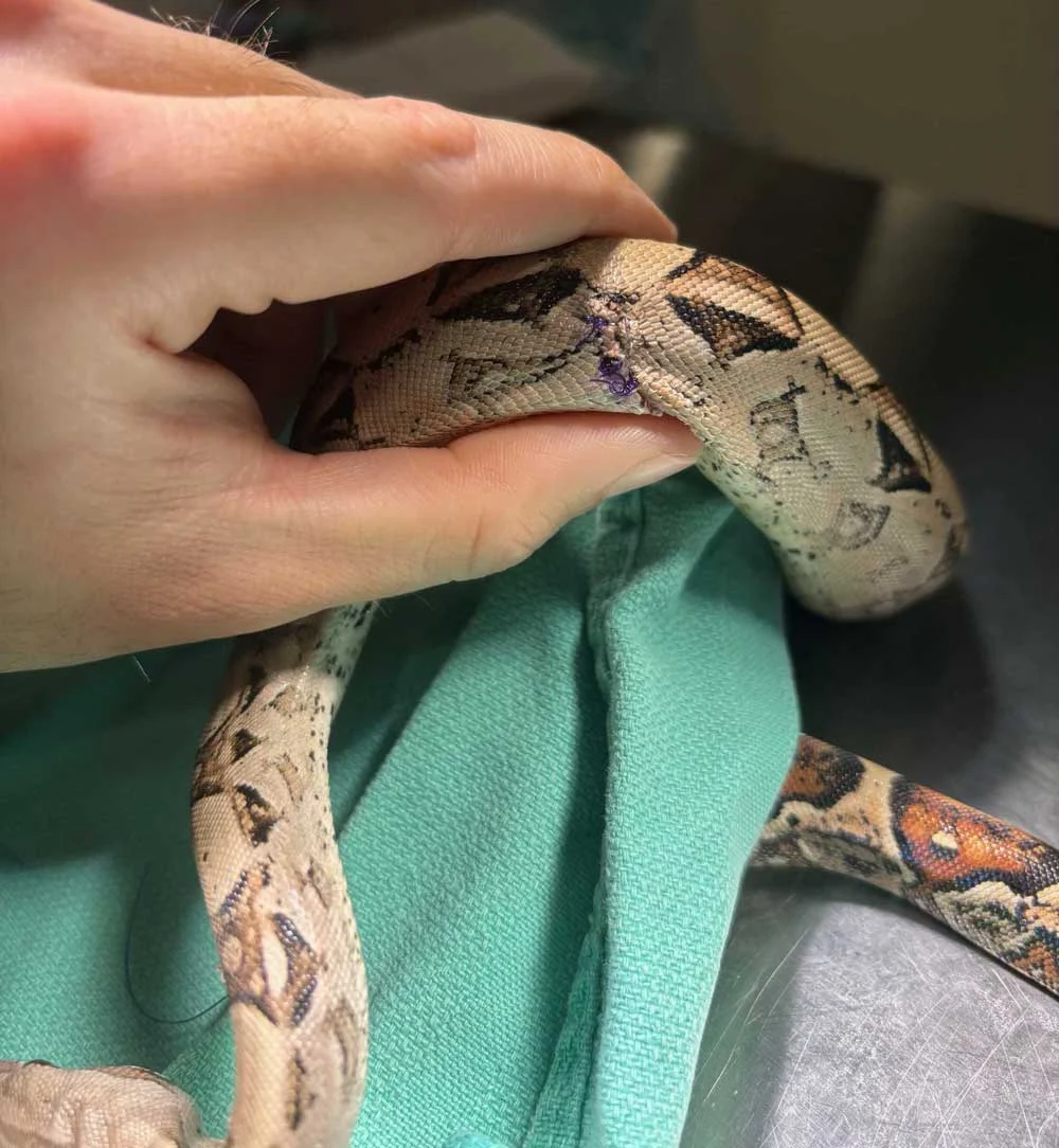
Trauma stems from both interior enclosure dynamics and handling accidents. Insecure decor can shift and crush a resting animal. Furthermore, excessive or improper handling of delicate species, such as giant day geckos, can result in "skin-sloughing" responses, where the animal sheds portions of its scales as an anti-predator defense. Rare mechanical failures, such as body wall hernias in leopard geckos, can also occur, causing tears in the internal musculature that allow visceral organs to push out beneath the skin.
3. Thermal Burns
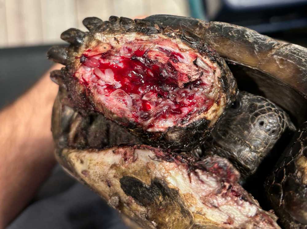
Thermal burns are entirely preventable injuries resulting from improper heat source management. The use of unregulated "heat rocks" (widely discouraged by modern herpetoculturists), heat mats lacking thermostats, or basking lamps that fail to cover the animal’s entire body frequently result in severe first- to third-degree burns. Large monitor lizards trying to warm their massive frames under an isolated, pinpoint bulb are particularly susceptible.
4. Traumatic Bites
Bites occur in communal housing setups or during feeding interactions. Overcrowded or improperly provisioned enclosures lacking adequate hides, thermal gradients, and food stations frequently spark aggressive territorial combat. Similarly, offering live mammalian prey or leaving unsupervised crickets in an enclosure overnight often results in severe defensive chewing injuries on the reptile’s skin.
5. Edema and Systemic Fluid Accumulation
Edema—the pathological pooling of fluid beneath the dermis—manifests as soft, compressible swellings. Rather than being a primary skin disorder, subcutaneous edema is typically a secondary indicator of severe internal disease, including congestive heart failure (CHF), advanced hepatic lipidosis (liver disease), or renal failure.
6. Neoplasia (Cancer)
Not every cutaneous lump is an abscess. Reptiles are susceptible to a wide array of malignancies. Veterinary oncologists have documented melanomas in aquatic turtles, squamous cell carcinomas, mast cell tumors, and sarcomas in bearded dragons, as well as benign lipomas in snakes.
7. Nutritional Deficiencies and Metabolic Disorders
Nutritional imbalances heavily impact epithelial tissues.
- Vitamin A Deficiency: Vitamin A is vital for maintaining healthy, moist mucous membranes, including the lining of eyelids, the oral cavity, and the hemipenes. Deficiencies trigger the accumulation of keratinous debris, leading to ocular blockages in leopard geckos, aural (ear) abscesses in aquatic turtles, hemipenal plugs in males, and stomatitis (infectious mouth rot).
- Gout: Articular gout occurs when uric acid crystals precipitate within joint capsules and soft tissues, causing excruciating pain and firm, swollen joints. Aspiration of these joints reveals characteristic chalky white debris. Management requires pain mitigation, urate-lowering medications like allopurinol, and strict dietary and hydration adjustments.
Official Responses: Veterinary Guidelines and Prevention Protocols
To address the high prevalence of preventable skin conditions, the Association of Reptile and Amphibian Veterinarians (ARAV) and clinical practitioners emphasize proactive husbandry protocols.
According to Dr. Eric Los Kamp, DVM, an exotic animal and wildlife veterinarian at Winter Park Veterinary Hospital, the cornerstone of dermatologic health lies in environmental management:
"The best way to minimize skin diseases in reptiles is providing optimal thermal and humidity gradients. Substrates must be cycled and replaced regularly; substrates that are too wet breed bacterial and fungal infections, while overly dry conditions precipitate dysecdysis. Furthermore, ensuring proper cleaning and maintenance of the enclosure is paramount. If there is a water feature and the reptile is aquatic, providing a powerful filter and frequent water changes will help reduce irritation to the skin from excessive waste in the water. Most importantly, if you see something concerning, bringing your animal to an exotic veterinarian immediately will help treat issues before they progress."
Implications: The Broader Impact on Reptile Welfare and Conservation
The prevalence of dermatologic diseases in captive reptiles carries significant implications for both the pet trade and wildlife conservation biology. In captivity, skin issues serve as a direct report card for husbandry standards. Because skin ailments are frequently the first visible symptom of a mismanaged environment, ignoring early signs of dysecdysis, mild dermatitis, or mite infestations often masks underlying systemic failures such as metabolic bone disease, chronic dehydration, or immunosuppression.
On a broader ecological scale, emerging dermatologic pathogens like Ophidiomyces ophiodiicola (snake fungal disease) and ranaviruses pose catastrophic threats to wild populations. As climate change shifts baseline environmental humidity and temperature ranges, wild reptiles face unprecedented physiological stressors that mirror the chronic conditions seen in poorly managed captive environments.
Ultimately, understanding the intricate biology of reptile scales and the vulnerabilities of their integumentary systems bridges the gap between better pet husbandry and the preservation of wild biodiversity. By treating the skin not merely as a surface covering, but as a dynamic reflection of overall health, keepers and veterinarians can vastly improve the quality of life for reptiles under human care.