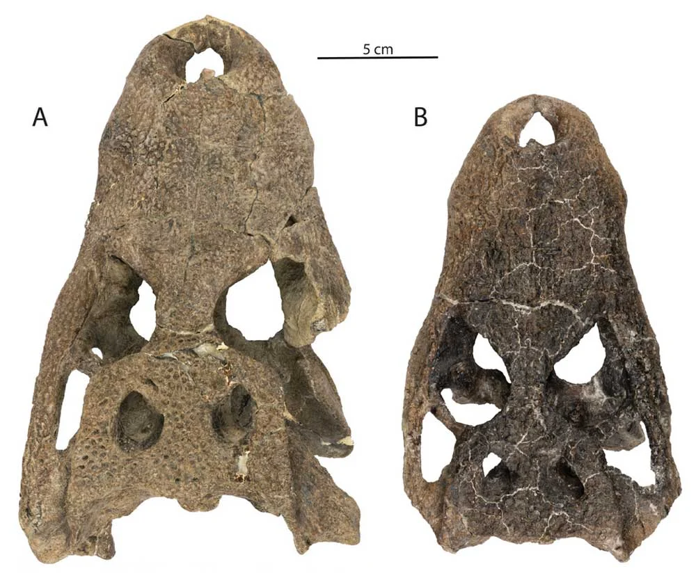
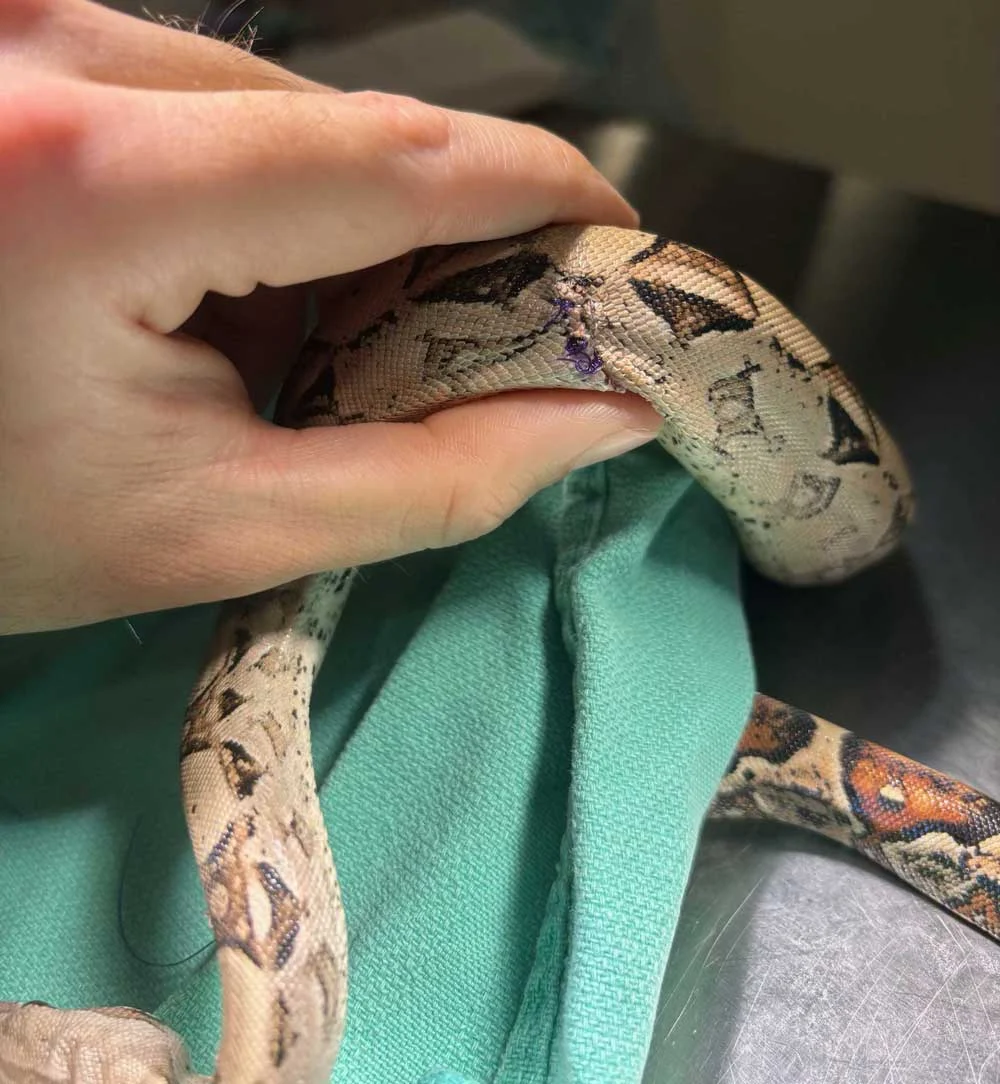
Reptilian scales are far more than static, decorative armor. These intricate dermal structures are evolutionary masterpieces that vary dramatically across taxa, playing critical roles in survival, movement, and physiological regulation. From the bony osteoderms embedded within the hides of varanids and crocodilians to the keeled texture of rattlesnakes, and the elastic skin of Uromastyx and chuckwallas designed to facilitate defensive inflation behaviors, reptilian integument is a dynamic organ. In species like snakes and certain geckos, specialized scales even fuse and clear to form the transparent spectacle that protects the eye, replacing traditional eyelids.
Because the skin is so thoroughly integrated into every facet of a reptile’s biology, any compromise to its integrity can trigger a cascading series of systemic health crises. Authored with insights from exotic animal practitioner Dr. Eric Los Kamp, DVM, of the Winter Park Veterinary Hospital, this comprehensive overview explores the multifaceted world of reptilian dermatology.
Disclaimer: This article is intended for educational purposes and is not designed to serve as a diagnostic or therapeutic manual. Any suspicion of a skin ailment in a pet reptile warrants immediate evaluation by a qualified exotic animal veterinarian.
The Anatomy of Vulnerability: Main Facts in Reptile Skin Health
The reptilian integumentary system acts as the primary barrier against environmental pathogens, mechanical trauma, and dehydration. However, the unique physiology of these animals means that skin diseases rarely remain localized. When a reptile’s skin is breached, damaged, or malfunctioning, it frequently serves as a window into broader systemic illnesses, husbandry deficiencies, or infectious loads within the environment.
Dermatologic conditions in captive reptiles generally fall into two broad categories: infectious and non-infectious. Understanding the etiology, clinical presentation, and underlying mechanisms of these conditions is paramount for keepers aiming to provide optimal husbandry and recognize early warning signs of distress.
Chronology of a Crisis: How Skin Issues Develop and Progress
Reptilian skin diseases typically follow distinct developmental timelines depending on whether the primary driver is environmental, infectious, or traumatic. Tracing the chronology of these conditions reveals why early intervention is so critical.
The Slippery Slope of Infectious Outbreaks
Infectious dermatologic diseases often begin with a microscopic breach in the skin barrier—often induced by micro-abrasions from abrasive substrates, low humidity, or ectoparasite bites.
- The Inoculation Phase: Pathogens (bacteria, fungi, or viral particles) colonize the superficial epidermal layers.
- The Proliferation Phase: If the reptile’s immune system is suppressed by suboptimal temperatures or stress, pathogens multiply rapidly. Bacterial dermatitis may manifest as localized ulceration, while fungal spores begin to digest keratin matrices.
- The Systemic Phase: Without intervention, localized infections breach the deeper dermal blood vessels, leading to systemic bacteremia, septicemia, or multi-organ failure. Conditions like Septic Cutaneous Ulcerative Dermatitis (SCUD) illustrate how localized skin trauma can rapidly escalate into a life-threatening, systemic emergency.
The Compound Effect of Environmental Stress
Non-infectious conditions, such as dysecdysis (abnormal shedding), follow a similarly progressive timeline rooted in husbandry failures:
- The Environmental Trigger: Humidity drops below species-specific requirements, or the thermal gradient is improperly calibrated.
- The Retention Phase: The normal enzymatic breakdown between the old and new epidermal layers fails. The shed is delayed, resulting in "stuck shed."
- The Constriction and Necrosis Phase: As the reptile continues to grow or subsequent shed cycles occur, retained layers accumulate. These layers act like tourniquets, particularly on the delicate digits and tails of lizards, restricting blood flow and culminating in dry gangrene and spontaneous amputation.
Supporting Data: Infectious Dermatologic Pathologies
Infectious agents exploit the unique structural properties of reptilian skin. Veterinary data highlights several key pathogens frequently encountered in clinical practice:
Bacterial Dermatitis and Abscesses
Bacterial skin infections manifest in two primary forms: diffuse dermatitis and localized abscesses. Bacterial dermatitis often presents with ulcerations (with or without hemorrhage), loss of normal scalation, crusting, and extreme pain upon palpation. Colloquially grouped under terms like "scale rot" or "tail rot," these infections involve various opportunistic gram-negative and gram-positive bacteria causing localized tissue necrosis.
A notable pathogen is Devrisea agamarum, a significant threat to Uromastyx and other agamid lizards. Epidemiological data underscores the insidious nature of this organism: bearded dragons frequently act as entirely asymptomatic carriers. They appear clinically normal while shedding the pathogen into collections, posing a severe contagion risk to more susceptible species.
Furthermore, reptile abscesses differ structurally from those found in mammals. Because reptiles lack peroxidase enzymes—the compounds responsible for liquefying pus—their abscesses produce a dense, caseous (cheesy) material. Because these thick-walled walls successfully wall off the infection from spreading, they also prevent systemic antibiotics from penetrating the core. Consequently, surgical debridement and complete excision are routinely required for resolution.
Viral Pathogens
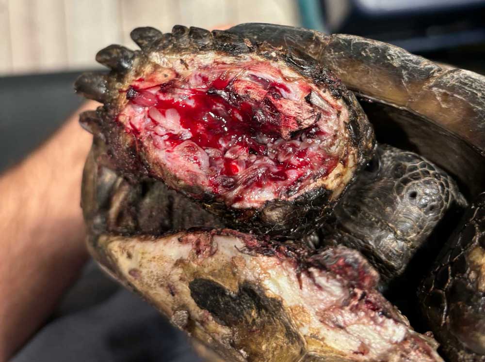
Viral etiologies frequently involve systemic infections that express themselves cutaneously. Poxviruses affect a wide range of reptilian species, mirroring the pathological behavior of mammalian poxviruses. In marine biology, the fibropapilloma virus—driven by a herpesvirus—creates massive, debilitating nodular growths on the faces and flippers of sea turtles. Ranaviruses and papillomaviruses are also documented drivers of reptile skin lesions.
Fungal Threats
Fungal dermatitis is notoriously contagious within captive collections and often carries systemic implications. High-profile examples include:
- Yellow Fungus Disease: Affects bearded dragons and other lizards, frequently proving fatal if it invades deep tissues.
- Snake Fungal Disease (Ophidiomyces ophiodiicola): Causes disfiguring facial crusting, scabs, and epidermal thickening in wild and captive ophidians.
- Emydomyces infections: Threaten semi-aquatic and box turtles, eroding keratinous shell scutes.
Ecto- and Endoparasites
Ectoparasites, most notably snake mites (Ophionyssus natricis), are prolific vectors for secondary bacterial and fungal infections while causing severe anemia through blood-feeding. Intriguingly, certain endoparasites (such as specific tapeworms and nematodes) utilize reptiles as intermediate hosts, forming dermal or subcutaneous nodules (granulomas). These parasites encyst within the reptile’s tissues, waiting for the animal to be consumed by a primary definitive host to complete their life cycle.
Supporting Data: Non-Infectious Dermatologic Pathologies
Non-infectious conditions are overwhelmingly tied to captive management errors, physiological imbalances, or genetic predispositions.
Trauma and Mechanical Injury
Trauma can occur both inside and outside the enclosure. Unsecured cage décor can shift or collapse, crushing or scraping animals. Furthermore, handling errors can result in acute damage; species such as giant day geckos possess fragile skin that easily sloughs off under excessive physical restraint or improper grip.
Rarer traumatic events include body wall hernias—documented in leopard geckos—where a tear in the underlying musculature allows internal organs to bulge subcutaneously.
Thermal Burns
Thermal burns remain a pervasive issue in reptile keeping. These injuries occur when keepers fail to provide properly regulated basking zones. Contributing factors include:
- Heat Rocks: Widely recognized by veterinary professionals as dangerous due to uneven heating elements and malfunctioning thermostats.
- Unregulated Heat Mats: Placed underneath tanks without a thermostat, often overheating and burning animals burrowing into the substrate.
- Under-Sized Basking Spots: Large species, such as monitor lizards, attempt to absorb heat from localized bulbs that only warm a fraction of their bodies, leading to compensatory positioning that results in localized burns.
Predatory and Conspecific Bites
Housing reptiles communally introduces the risk of conspecific aggression. Without adequate resources—such as multiple basking sites, visual barriers, and isolated feeding stations—dominant animals will inflict bite wounds on cage mates. Similar trauma occurs when live prey items (such as crickets or rodents) are left unattended in an enclosure and retaliate, chewing on a sleeping or docile reptile’s skin.
Systemic and Metabolic Manifestations: Edema, Cancer, and Nutrition
The skin also reflects internal organ failure and metabolic imbalance:
- Edema: Subcutaneous fluid accumulation presents as compressible, fluid-filled swellings and is frequently secondary to congestive heart failure (CHF), advanced renal disease, or hepatic lipidosis.
- Neoplasia: Masses are not always abscesses. Reptiles develop various malignancies, including melanomas in turtles, squamous cell carcinomas, mast cell tumors, and sarcomas in bearded dragons, as well as benign lipomas in snakes.
- Nutritional Deficiencies: Vitamin A is vital for maintaining healthy, moist mucous membranes. Deficiencies lead to keratinous debris accumulation, manifesting as eye caps in leopard geckos, aural abscesses in aquatic turtles, hemipenal plugs, and stomatitis (mouth rot).
- Articular Gout: Uric acid crystal deposition around joints causes intense pain and swelling. Aspiration reveals chalky white debris, managed through a combination of allopurinol, analgesics, and strict dietary and hydration adjustments.
Official Recommendations and Veterinary Protocols
To combat the rising incidence of preventable dermatologic disease, veterinary organizations and exotic specialists emphasize standardized husbandry protocols:
- Environmental Optimization: Maintain strict, species-appropriate thermal and humidity gradients. Utilize digital hygrometers and thermostats for all heating elements.
- Substrate Management: Cycle and replace substrates regularly. Excessively wet substrates invite bacterial and fungal blooms, while overly dry environments trigger shedding failures.
- Aquatic Hygiene: For aquatic and semi-aquatic species, install high-capacity filtration systems and perform frequent water changes to prevent waste accumulation from irritating delicate skin and plastrons.
- Immediate Veterinary Consultation: Experts stress that self-diagnosis or over-the-counter treatments often delay critical care. Prompt presentation to an ARAV (Association of Reptile and Amphibian Veterinarians) member veterinarian provides the best prognosis for recovery.
Implications for the Future of Reptile Husbandry
The state of a reptile’s skin is a direct report card on its captive environment and internal physiological health. As veterinary medicine advances, the understanding of how systemic diseases manifest cutaneously continues to grow.
For the reptile-keeping community, the implication is clear: proactive husbandry is vastly superior to reactive medicine. By prioritizing environmental precision, nutritional balance, and biosecurity—especially regarding asymptomatic carriers of pathogens like Devrisea agamarum—keepers can dramatically reduce the prevalence of dermatologic disease. Ultimately, safeguarding the health of reptilian scales ensures the well-being of the entire animal, preserving the evolutionary marvels that make these creatures so captivating.