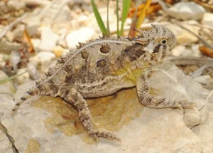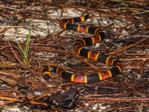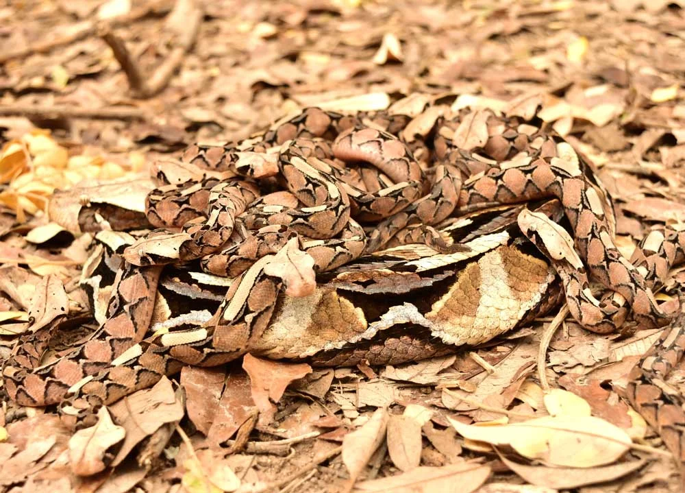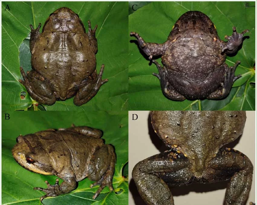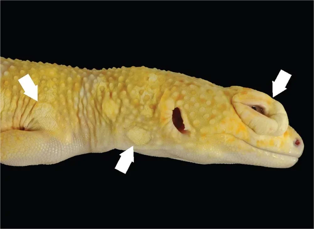
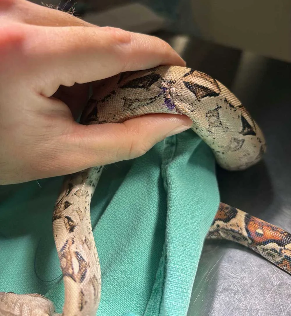
Reptile scales are far more than static, decorative armor. Far from being uniform coverings, these complex integumentary systems vary radically across taxa, serving as dynamic evolutionary masterpieces tailored to specific ecological niches. Varanids (monitor lizards) and crocodilians feature osteoderms—actual bones embedded directly into their scales—providing formidable natural defense. Rattlesnakes boast keeled scales with a raised ridge down the center, while desert-dwellers like Uromastyx and chuckwallas possess elastic hides that accommodate drastic bodily inflation behaviors. In numerous gecko species and all snakes, the eyelids have fused and transparently evolved into a protective scale known as the spectacle.
Because the skin is so fundamentally intertwined with every aspect of a reptile’s physiology, thermoregulation, and defense, dermatologic issues rarely remain localized. Skin pathology often serves as a flashing neon warning sign of broader systemic distress, improper husbandry, or infectious invasion.
Disclaimer: This article provides a broad educational overview of conditions affecting reptile skin and is not intended to serve as a diagnostic or therapeutic manual. Any pet owner observing skin abnormalities should seek immediate evaluation from a qualified reptile veterinarian.
Infectious Dermatologic Diseases
Infectious agents—ranging from microscopic bacteria and aggressive fungi to stealthy viruses and stubborn parasites—pose a constant threat to the integumentary integrity of captive and wild reptiles alike.
Bacterial Pathogens and Abscess Formation
Bacterial skin infections manifest in two primary clinical presentations: diffuse bacterial dermatitis and localized abscesses. Bacterial dermatitis can ravage an animal’s skin, presenting as weeping ulcerations with or without hemorrhage, scalation loss, crusting, and extreme localized pain upon palpation.
A prime clinical example is Devrisea agamarum, a significant and highly contagious pathogen affecting Uromastyx and other agamid lizards. Alarmingly, popular pets like bearded dragons often act as asymptomatic carriers, appearing 100% clinically normal while actively shedding the pathogen to far more susceptible species. Other generalized cutaneous infections, colloquially grouped under terms like “rot” (e.g., tail rot or scale rot), involve progressive localized tissue necrosis driven by opportunistic bacteria. Furthermore, severe bacterial dermatitis can act as a cutaneous manifestation of grave systemic illnesses, such as Septic Cutaneous Ulcerative Dermatitis (SCUD).
Unlike mammals, reptiles completely lack peroxidase enzymes—the chemical agents responsible for liquefying pus. Consequently, reptile abscesses do not form fluid-filled pockets; instead, they develop dense, caseous, cheese-like cores encased in thick fibrous walls. These thick walls, while evolutionary mechanisms designed to prevent a systemic spread of infection, also block systemic antibiotics from penetrating the core. Thus, treating reptile abscesses almost universally requires surgical debridement and excision by an exotic veterinarian.
Viral Threats Across Taxa
Viral etiologies are increasingly recognized in reptile medicine, frequently launching attacks that manifest simultaneously on the skin and within internal organs. Poxviruses routinely afflict multiple reptile species, mirroring the pathogenic behaviors of their mammalian counterparts.
In marine conservation, the fibropapilloma virus—driven by a herpesvirus—wreaks havoc on sea turtle populations by generating massive, debilitating tumor-like nodules across the face, eyes, and flippers, severely impairing vision and swimming capabilities. Ranaviruses and papillomaviruses are also documented culprits behind disfiguring and fatal dermal lesions.
Fungal Infections and High Contagiosity
Fungal dermatitis in reptiles is notoriously aggressive. Fungal organisms frequently transition from superficial skin invaders to systemic pathogens capable of shutting down vital organs. Compounding the danger, these infections are exceptionally contagious within multi-reptile collections or poorly sanitized setups.
High-profile fungal threats include Yellow Fungus Disease (caused by Nannizziopsis species) which devastates bearded dragons and other captive lizards, Snake Fungal Disease (Ophidiomyces ophiodiicola) which threatens wild and captive snake populations alike, and Emydomyces species, which target the shells and skin of box turtles and aquatic pond turtles.
Ecto- and Endoparasites
Parasitic burdens can be divided into external menaces and internal hitchhikers. Ectoparasites, most notably snake mites (Ophionyssus natricis), are a chronic scourge in captivity. These microscopic vampires burrow between scales to feed on blood, causing severe anemia, irritation, and secondary bacterial or fungal infections as the reptile frantically rubs against enclosure decor to find relief.
Meanwhile, certain endoparasites—such as specific tapeworms and nematodes—can trigger localized nodules or granulomas beneath the skin. This occurs when a reptile serves as an intermediate or accidental host. The parasite encysts in the musculature or subcutis, waiting for the reptile to be consumed by a primary predator-host, thereby completing the parasite’s complex life cycle.
Non-Infectious Dermatologic Diseases
While pathogens capture significant veterinary attention, a vast majority of reptile skin cases stem from environmental mismanagement, traumatic incidents, nutritional deficiencies, and internal organ failure.
Dysecdysis: The Mechanics of Abnormal Shedding
Ecdysis, or shedding, is a tightly regulated physiological process. When disrupted, it results in dysecdysis, or abnormal shedding. Most commonly, keepers encounter delayed shedding, leading to "stuck shed." This is almost universally rooted in suboptimal husbandry, primarily chronic low ambient humidity or improper hydration.
In mild cases, tepid water soaks and gentle manual assistance can alleviate retained skin. However, if multiple layers of stuck shed are allowed to accumulate over successive cycles, they act as a constrictive band. This cuts off micro-circulation, leading directly to ischemic necrosis and auto-amputation of digits and tails—a pathology frequently observed in leopard geckos and arboreal lizards. Conversely, dysecdysis can also present as accelerated shedding, triggered by chronic dermal irritation from mites, surgical sutures, or severe metabolic imbalances.
Trauma and Mechanical Injury
Trauma can occur both inside and outside the enclosure. Within the habitat, poorly secured rock structures, unstable branches, or abrasive mesh can cause severe lacerations and abrasions. Out-of-enclosure handling demands vigilance; fragile species like giant day geckos possess specialized defensive skin adaptations allowing them to readily slough off portions of skin and scales when seized by a predator (or an overly aggressive human handler).
More unusual traumatic presentations include body wall hernias, occasionally documented in leopard geckos. These tears in the underlying musculature allow internal abdominal organs to herniate and push outward beneath the skin envelope.
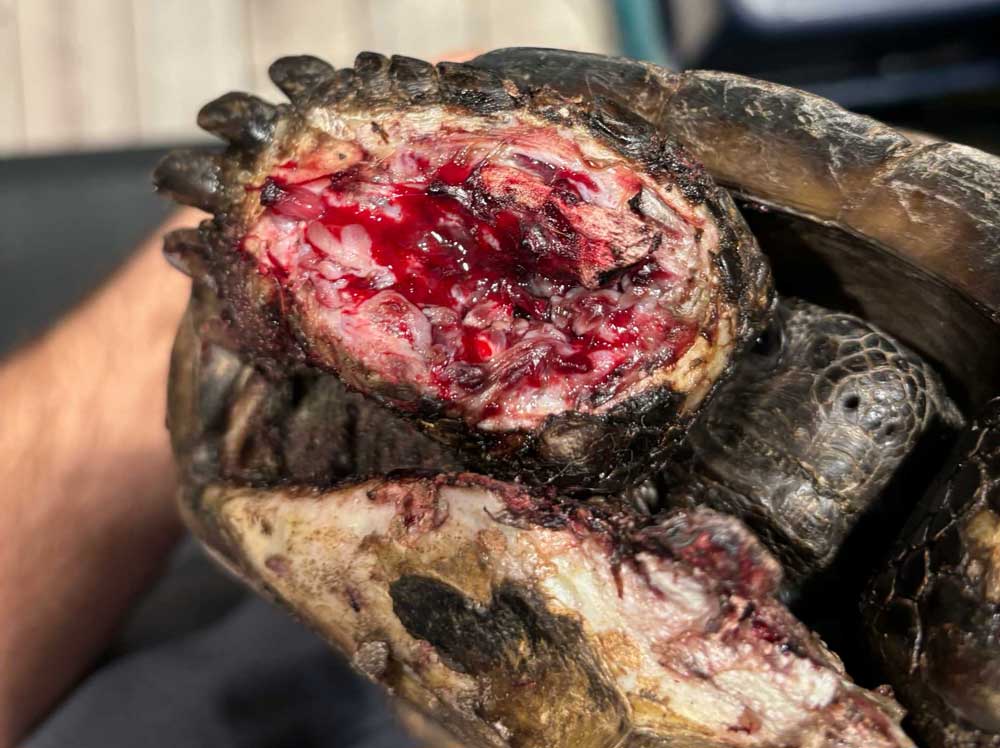
Thermal Burns
Thermal burns are an entirely preventable yet tragically common emergency in reptile husbandry. They occur when animals are denied proper thermal gradients or exposed to unsafe heating elements.
The use of commercial "heat rocks"—unregulated electrical heating stones—remains a major hazard and is strongly condemned by exotic veterinarians. Similarly, utilizing undertank heating pads without a digital thermostat or providing a basking spot too physically small for the animal (such as a massive monitor lizard attempting to absorb heat from a single concentrated spotlight) frequently results in severe, deep-tissue thermal burns. Prevention relies exclusively on utilizing high-quality proportional thermostats and constructing broad, evenly distributed basking zones capable of warming the animal’s entire anatomical mass.
Traumatic Bites
Bites occur in two primary contexts: cohabitating tank mates and aggressive prey items. Communal housing of solitary reptile species invites territorial aggression, resulting in severe lacerations, missing tails, and bite-induced systemic infections. If cohabitation is attempted under expert supervision, keepers must supply multiple independent basking zones, visual hides, and feeding stations to minimize conflict.
Prey-induced trauma is equally devastating. Offering live rodents to snakes frequently results in the rodent defending itself by chewing horrific, deep wounds into the snake’s flesh. Similarly, leaving uneaten crickets loose in a lizard enclosure overnight often leads to the insects chewing on the sleeping reptile’s scales and digits.
Edema and Fluid Imbalance
Edema—the pathological accumulation of serous fluid within interstitial spaces under the skin—presents as soft, compressible, fluid-filled swellings. Far from being a primary skin disease, subcutaneous edema is frequently a secondary symptom pointing to profound internal organ failure, including congestive heart failure (CHF), advanced liver disease, or terminal renal failure.
Neoplasia (Cancer)
Not every subcutaneous mass is a caseous abscess or a cyst; reptiles are fully susceptible to neoplastic transformation. Veterinary oncologists have documented a wide array of cancers in reptilian patients, including:
- Melanomas in aquatic turtles
- Squamous cell carcinomas, mast cell tumors, and sarcomas in bearded dragons
- Lipomas (benign fat tumors) in various snake species
Nutritional Dermatopathies and Metabolic Disorders
Nutrition plays a foundational role in integumentary health. Vitamin A is critical for maintaining healthy, moist mucous membranes, including the epithelial linings of the mouth, eyelids, and hemipenes. Dietary deficiencies lead to keratinous debris accumulation. In leopard geckos, this causes debilitating ocular keratosis; in aquatic turtles, it drives chronic aural (ear) abscesses; in lizards, it triggers hemipenal plugs and stomatitis (mouth rot).
Furthermore, systemic metabolic conditions like articular gout can manifest via visible, painful joint swellings. When aspirated by a veterinarian, these joints yield a chalky white accumulation of uric acid crystals. Managing gout requires a multi-pronged approach involving targeted pain management, allopurinol to decrease uric acid synthesis, and aggressive dietary and hydration overhauls.
Chronology of Care: What Happens During a Veterinary Workup?
When an owner brings a reptile with a suspected dermatologic issue to an exotic animal hospital, the clinical timeline generally follows a standardized diagnostic chronology:
- Husbandry Audit (Initial Consultation): The veterinarian reviews ambient temperatures, hot-spot gradients, hygrometer accuracy, diet, supplementation schedules, and substrate composition. Many skin issues are immediate reflections of environmental error.
- Physical and Dermatologic Examination: The clinician examines the entire body under magnification, evaluating scale integrity, looking for mites, checking spectacles, and palpating for abnormal masses or localized pain.
- Diagnostic Sampling: Depending on the presentation, the vet may perform skin scrapings (for mites or fungal spores), fungal/bacterial cultures and sensitivities, fine-needle aspirations of masses or swellings, or blood work to assess internal organ function (liver/kidneys).
- Targeted Intervention: Treatment plans are established, which may range from environmental adjustments and medicated soaks to systemic antibiotic/antifungal therapy, parasite eradication protocols, or surgical debridement of necrotic tissue and abscesses.
Supporting Data & Epidemiological Insights
- Husbandry Correlation: Clinical data from exotic veterinary practices indicates that over 70% of mild-to-moderate reptile skin diseases (such as stuck shed and superficial dermatitis) are directly traceable to improper humidity levels or unhygienic substrate maintenance.
- Contagiosity Rates: Fungal pathogens like Ophidiomyces ophiodiicola and Nannizziopsis species exhibit near 100% transmission rates in shared-air or multi-reptile facilities if strict quarantine and biosecurity protocols are ignored.
- Anatomical Vulnerability: The distal extremities (toes and tail tips) account for an estimated 85% of auto-amputations resulting from chronic dysecdysis due to constrictive layers of retained shed.
Official Responses and Veterinary Guidance
The Association of Reptile and Amphibian Veterinarians (ARAV) strongly emphasizes proactive husbandry over reactive medical treatment. According to clinical guidelines issued by exotic specialists, the vast majority of dermatologic emergencies can be successfully prevented through meticulous environmental control.
"The reptile integument is a direct mirror of its internal health and environmental stability," notes veterinary literature from leading reptile specialists. "When a keeper notices a change in scalation, color, or texture, the clock is already ticking on a potential systemic cascade. Early intervention by a qualified exotic veterinarian is the single greatest factor in ensuring a positive clinical outcome."
Implications for Reptile Keepers and Conservation
The intersection of skin health and reptile husbandry carries profound implications for both domestic pet owners and global wildlife conservationists.
For the average pet keeper, understanding the etiology of skin disease shifts the paradigm from passive observation to active environmental management. Maintaining pristine substrates, investing in digital hygrometers and thermostats, and eliminating live feeding hazards save animals from agonizing, preventable conditions.
On a broader ecological scale, understanding reptile skin pathogens—such as the relentless spread of Snake Fungal Disease and sea turtle fibropapillomatosis—informs conservation strategies for vulnerable wild populations. As climate change shifts ambient moisture and temperature profiles in natural habitats, wild reptiles face unprecedented environmental stressors that directly compromise their natural immunological and cutaneous defenses. Recognizing skin as a complex, dynamic organ rather than mere physical wrapping allows both veterinarians and keepers to better protect these remarkable animals, ensuring their resilience for generations to come.